Microtubule Facts For Kids
Microtubules are polymers of tubulin that form part of the cytoskeleton, providing structure and shape to eukaryotic cells.


Do more with AI
Introduction
Microtubules are tiny tubes found inside our cells! 🌟They are made of special proteins called tubulin. Picture them as the building blocks that help give cells their shape and structure, just like how walls support a house! Microtubules are important for many cell activities, and they help cells move and stay stable. They are found in all living things with complex cells, called eukaryotes. This includes plants, animals, and even humans! Without microtubules, cells would be floppy and not work properly. That’s why they are super important for life! 🦠
Images of Microtubule
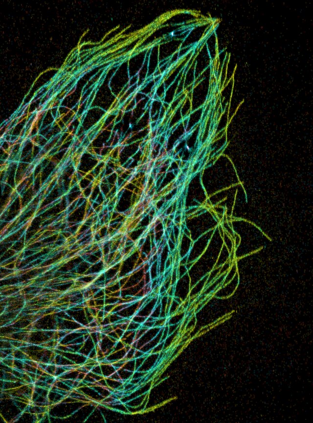
Microtubules are one of the cytoskeletal filament systems in eukaryotic cells. The microtubule cytoskeleton is involved in the transport of material within cells, carried out by motor proteins that move on the surface of the microtubule.
![Cartoon representation of the structure of α(yellow)/β(red)-tubulin heterodimer, GTP and GDP.[11]](https://upload.wikimedia.org/wikipedia/commons/thumb/d/dc/Tubulin_dimer_1JFF.png/500px-Tubulin_dimer_1JFF.png)
Cartoon representation of the structure of α(yellow)/β(red)-tubulin heterodimer, GTP and GDP.[11]

Components of the eukaryotic cytoskeleton include: nuclei (blue), microtubules (green), and actin filaments (red).

Image of a fibroblast cell containing fluorescently labeled actin (red) and microtubules (green).

A cytoplasmic dynein motor bound to a microtubule.

A kinesin molecule bound to a microtubule.

A 3D diagram of a centriole. Each circle represents one microtubule. In total there are 27 microtubules organized into 9 bundles of 3.

This diagram depicts the organization of a typical mitotic spindle found in animal cells. Shown here are the three main types of microtubules during mitosis and how they are oriented in the cell and the mitotic spindle.
Microtubules are one of the cytoskeletal filament systems in eukaryotic cells. The microtubule cytoskeleton is involved in the transport of material within cells, carried out by motor proteins that move on the surface of the microtubule.
Cartoon representation of the structure of α(yellow)/β(red)-tubulin heterodimer, GTP and GDP.[11]
Components of the eukaryotic cytoskeleton include: nuclei (blue), microtubules (green), and actin filaments (red).
Image of a fibroblast cell containing fluorescently labeled actin (red) and microtubules (green).
A cytoplasmic dynein motor bound to a microtubule.
A kinesin molecule bound to a microtubule.
A 3D diagram of a centriole. Each circle represents one microtubule. In total there are 27 microtubules organized into 9 bundles of 3.
This diagram depicts the organization of a typical mitotic spindle found in animal cells. Shown here are the three main types of microtubules during mitosis and how they are oriented in the cell and the mitotic spindle.
Structure Of Microtubules
Microtubules are like little straw-shaped tubes that can change shape! 🌈They are made from two types of proteins called alpha and beta tubulin. When these proteins stick together, they create long chains that twist and form a tube. Each tube is about 25 nanometers wide, which is really, really tiny—like a million times thinner than a human hair! Microtubules can grow longer or shorten, and they can connect with each other to make networks. They are sturdy but flexible, allowing cells to keep their shape while still moving around! 🏗️
Functions Of Microtubules In Cells
Microtubules help cells do many important things! 🌲First, they provide support and shape to the cell, keeping it strong. Second, they act like train tracks for tiny cargoes that are carried around by motor proteins. These motor proteins can move things like nutrients and other important materials across the cell. Third, microtubules help separate chromosomes during cell division, making sure each new cell gets the right number of chromosomes. Microtubules are essential to keep everything running smoothly in the cell! ⚙️
Microtubules In Cilia And Flagella
Cilia and flagella are tiny hair-like structures that help some cells move! 🏊♂️ Cilia are short and can be found on cells like the ones in our throat, helping to move mucus out. Flagella are longer and usually found on sperm cells, helping them swim toward an egg! Both cilia and flagella have a special arrangement of microtubules inside them, usually in a “9 + 2” pattern. This means there are nine pairs of microtubules around two single ones in the center. This design helps them wave back and forth to create movement! 💨
Microtubules And Cellular Transport
Microtubules are like delivery trucks in a city, helping move important things around the cell! 🚚Motor proteins, like kinesin and dynein, use microtubules as tracks to transport materials such as proteins, organelles, and waste. Kinesin moves cargo away from the center of the cell, while dynein moves cargo back toward the center. This transportation helps cells communicate and perform their tasks. Imagine a busy city with delivery trucks! Microtubules make sure all the parts of the cell stay connected and get to where they need to go quickly! 🏙️
Role Of Microtubules In Cell Division
Microtubules play a key role during cell division (when one cell divides into two!). 🎉During this process, called mitosis, microtubules form a special structure called the mitotic spindle. This spindle helps pull apart chromosomes, which are like tiny instruction manuals for the cell. Each new cell needs to get the right instructions to function properly! Microtubules stretch across the cell like ropes, making sure that both new cells get equal numbers of chromosomes. Without microtubules, cells wouldn't be able to divide correctly, which is super important for growth and repair! 🔧
Microtubule-associated Proteins (maps)
Microtubule-Associated Proteins (MAPs) are special helpers that work with microtubules! 🦸♂️ These proteins help stabilize microtubules, making them stronger and less likely to break. Some MAPs also help organize microtubules into structures called bundles, which can aid in moving things around! Other MAPs can help control how fast microtubules grow or shrink. They are like managers in a workplace, guiding microtubules so that they function well and help the cell do its job. Without these proteins, microtubules wouldn't be able to work effectively! 📋
Diseases Related To Microtubule Dysfunction
Sometimes, things can go wrong with microtubules, leading to diseases! 😷For example, when microtubules don't work properly, it can lead to issues like cancer or neurodegenerative diseases, such as Alzheimer's. In cancer, changes in microtubules can make cells divide uncontrollably. In neurodegenerative diseases, problems with microtubules can affect how brain cells communicate. Scientists study these problems to find ways to help people feel better. Learning about microtubules and their functions can help us understand and fight these diseases! 🧬
Applications Of Microtubule Study In Medicine
Studying microtubules helps improve medicine and treatments! 💉For instance, certain cancer drugs target microtubules to stop cancer cells from dividing, making them less able to grow. These drugs are called “chemotherapeutics.” Knowing how microtubules function helps doctors understand diseases and find new ways to treat them. Researchers are also exploring how to use microtubules in biotechnology, like making advanced medicines or using them to deliver drugs to specific parts of the body. Microtubules are truly remarkable and have important roles in keeping us healthy! 😊
Research Techniques For Studying Microtubules
Scientists use different techniques to study microtubules and understand their functions better! 🔬One popular method is using microscopy, which allows them to see microtubules up close. They can tag microtubules with special dyes that glow under a microscope. Other methods include using electron microscopes, which can show even more detail! Scientists also study how microtubules change during cell division or when carrying cargo. By understanding how microtubules work, they can learn how to fix problems when cells don't work right! 📊
Microtubule Dynamics: Polymerization And Depolymerization
Microtubules are very dynamic, which means they can change their size and shape! ⚡When they are made longer, we call it "polymerization." This is when tubulin proteins stick together to form a longer tube. On the other hand, "depolymerization" happens when the tubulin proteins come apart, making the tube shorter. This ability to grow and shrink at fast speeds is super important for how cells divide and move. Just like a magician pulls a scarf from their sleeve, microtubules can quickly change their length to help the cell! 🎩
Microtubule Quiz
Learn more about Microtubule
