Computed Tomography Facts For Kids
Computed Tomography (CT) is a medical imaging technique that combines multiple X-ray images to create detailed internal pictures of the body, helping doctors diagnose various health issues.


Do more with AI
Introduction
Computed Tomography, or CT scans for short, are special machines that take pictures of the inside of our bodies! 📸🩺 They use X-rays to create detailed images that help doctors see bones, organs, and tissues. Imagine a giant donut-shaped camera that spins around you while you lie still inside! This technology is super helpful in diagnosing illnesses and injuries. The first CT scans were used in the 1970s, and now they’re found in hospitals all around the world. 🌍With CT scans, doctors can quickly find out what's happening inside without needing to perform surgery. Isn't that cool?
Images of Computed Tomography
Drawing of CT fan beam and patient in a CT imaging system

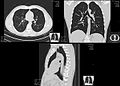
CT scan of the thorax. The axial slice (right) is the image that corresponds to number 2/33 on the coronal slice (left).

CT Perfusion scan of the brain

PET-CT scan of chest

Computed tomography of human brain, from base of the skull to top. Taken with intravenous contrast medium. Commons: Scrollable computed tomography images of a normal brain
HRCT images of a normal thorax in axial, coronal and sagittal planes, respectively. Click here to scroll through the image stacks.

Bronchial wall thickness (T) and diameter of the bronchus (D)

Example of a CTPA, demonstrating a saddle embolus (dark horizontal line) occluding the pulmonary arteries (bright white triangle)

CT scan of a normal abdomen and pelvis, in sagittal plane, coronal and axial planes, respectively. Click here to scroll through the image stacks.

Types of presentations of CT scans: − Average intensity projection− Maximum intensity projection− Thin slice (median plane)− Volume rendering by high and low threshold for radiodensity
Drawing of CT fan beam and patient in a CT imaging system
CT scan of the thorax. The axial slice (right) is the image that corresponds to number 2/33 on the coronal slice (left).
CT Perfusion scan of the brain
PET-CT scan of chest
Computed tomography of human brain, from base of the skull to top. Taken with intravenous contrast medium. Commons: Scrollable computed tomography images of a normal brain
HRCT images of a normal thorax in axial, coronal and sagittal planes, respectively. Click here to scroll through the image stacks.
Bronchial wall thickness (T) and diameter of the bronchus (D)
Example of a CTPA, demonstrating a saddle embolus (dark horizontal line) occluding the pulmonary arteries (bright white triangle)
CT scan of a normal abdomen and pelvis, in sagittal plane, coronal and axial planes, respectively. Click here to scroll through the image stacks.
Types of presentations of CT scans: − Average intensity projection− Maximum intensity projection− Thin slice (median plane)− Volume rendering by high and low threshold for radiodensity
Preparing For A Ct Scan
If you need a CT scan, preparing for it is simple! 🛏️ First, your doctor will explain everything to you and your parents. They might ask you to wear a patient gown for comfort. 👗Next, you might need to avoid eating for a few hours before the scan, depending on what part of your body they’re scanning. You’ll lie on a bed that moves into the CT scanner, and the staff will be there to help you feel comfortable. 😌Remember to stay still during the scan, but don’t worry! It’s quick, and soon you’ll be on your way! 🌈
Applications In Medicine
CT scans are super important for doctors! They help in finding broken bones, tumors, and other health issues. 🏥For instance, if you hurt your leg, a CT scan can show if there are any fractures. It can also help detect diseases like cancer and infections. 🦠In emergency rooms, CT scans help doctors quickly assess what’s wrong with patients after accidents. They make diagnosis faster, which means treatment can start sooner. 💪CT technology is also useful during surgeries, guiding doctors to precisely where they need to work. So, CT scans play a big role in keeping us healthy! 🌟
How Computed Tomography Works
A CT scan machine looks like a big donut. 🌈When you're ready, you lie down on a bed that moves through the donut. Inside, an X-ray tube spins in a circle. It takes lots of pictures from different angles while the bed slowly moves. ⏲️ These pictures are then sent to a computer that combines them to create detailed images of your insides! The doctor can then see everything in a cross-section, like slicing a loaf of bread! 🍞The process only takes a few minutes, and it provides clear images to help doctors understand any problems you might have.
History Of Computed Tomography
In the 1970s, two brilliant inventors, Sir Godfrey Hounsfield from the UK and Dr. Allan Cormack from South Africa, created the first CT scan. 🔍They teamed up to make technology that could take images of the human body without cutting it open! Their work changed medicine forever! In 1979, they received the Nobel Prize for their amazing invention. 🎉The first CT scanner was installed in a London hospital and quickly spread worldwide. By the 1980s, many hospitals had CT scanners, making it easier for doctors to help patients. Today, CT scans are a vital part of modern medicine!
Safety And Risk Considerations
Safety is very important when it comes to CT scans! While they are generally safe, it’s good to know some things. ⚠️ CT scans use a special kind of radiation, similar to what you’d get in everyday life. Still, doctors try to avoid unnecessary scans to limit exposure. For children, special care is taken to ensure their safety, as they are more sensitive to radiation. 👶Pregnant women should discuss with their doctors if a CT scan is necessary. 🗣️ In most cases, the benefits of a scan far outweigh the risks, helping doctors diagnose issues effectively!
Types Of Computed Tomography Scans
There are different types of CT scans, each used for different parts of the body! 🦴For example, a "CT scan of the abdomen" looks at your stomach and intestines, while a "CT scan of the brain" helps doctors see the head and brain. 🧠There's also a "CT angiography," which shows blood vessels and helps find blockages. Some scans use special dyes, called contrast agents, to highlight particular organs. 💉No matter the type, each scan gives doctors important information to keep us healthy! ❤️ Understanding which scan to use can help doctors make the best decisions for their patients.
Common Conditions Diagnosed With Ct
CT scans can help diagnose many conditions! 🚑They are often used for finding broken bones or fractures, which is super important in emergencies. 🦴Doctors also use CT scans to identify tumors, cysts, and infections. For instance, when someone has abdominal pain, a CT scan can help check for appendicitis. 🌟Other conditions like lung diseases and brain injuries can also be explored with CT. It’s like having a super detective in the hospital that helps doctors find out what’s going on inside our bodies! 🕵️♂️ CT scans truly make it easier to understand health!
Benefits And Limitations Of Ct Scans
CT scans have many benefits! They provide detailed images quickly and are often painless. 🌈They help doctors make accurate diagnoses and find problems easily, which can save lives! However, there are some limitations. CT scans use X-rays, which means there is a small amount of radiation exposure. 😟This is usually safe for most people, but doctors try to limit unnecessary scans. Another limitation is that there may be concerns for young children or pregnant women. However, the benefits of helping diagnose health conditions often outweigh the risks, as doctors take precautions.
Future Developments In Ct Technology
The future of CT scans is exciting! 🚀Scientists are always working to make them better and safer. New technologies are being developed to reduce radiation doses while still getting clear images. 💡One advancement is artificial intelligence, which can help doctors analyze images faster and more accurately! AI can assist in spotting issues that might be missed. 👀Additionally, researchers are exploring portable CT machines, which could help in emergencies or places without big machines! ❤️ With these advancements, CT technology will continue to improve patient care and make medical imaging even more efficient!
Comparison With Other Imaging Techniques
CT scans are just one way to take pictures of our insides. 📷There are others like X-rays, MRIs, and ultrasounds. X-rays give a quick look at bones but don’t show organs very well. 🦴MRIs, on the other hand, use magnets to create detailed pictures of soft tissues, but they take longer and may be noisier! 🤯Finally, ultrasounds use sound waves to visualize organs, especially during pregnancy. 🤰In comparison, CT scans are great for quickly seeing both bones and organs in one scan, making them a popular choice for doctors everywhere! 🌍
Computed Tomography Quiz
Learn more about Computed Tomography
