Large Intestine Facts For Kids
The large intestine is the final part of the digestive system, responsible for absorbing water and forming waste material for elimination.


Do more with AI
Introduction
The large intestine is a super important part of our body! 🏃♂️ It’s like a big tube, about 5 feet long, located at the end of the digestive system. You can find it below your stomach and small intestine. The large intestine is shaped like an upside-down "U" and connects to the rectum, where our body gets rid of waste! 💩It has four main parts: the cecum, colon, rectum, and anal canal. The large intestine helps turn leftover food into poop, which is very important for keeping our bodies healthy!
Images of Large Intestine
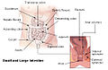
Illustration of the large intestine.

Inner diameters of colon sections

3D file generated from computed tomography of large intestine
![Colonic crypts (intestinal glands) within four tissue sections. The cells have been stained to show a brown-orange color if the cells produce the mitochondrial protein cytochrome c oxidase subunit I (CCOI), and the nuclei of the cells (located at the outer edges of the cells lining the walls of the crypts) are stained blue-gray with haematoxylin. Panels A, B were cut across the long axes of the crypts and panels C, D were cut parallel to the long axes of the crypts. In panel A the bar shows 100 μm and allows an estimate of the frequency of crypts in the colonic epithelium. Panel B includes three crypts in cross-section, each with one segment deficient for CCOI expression and at least one crypt, on the right side, undergoing fission into two crypts. Panel C shows, on the left side, a crypt fissioning into two crypts. Panel D shows typical small clusters of two and three CCOI deficient crypts (the bar shows 50 μm). The images were made from original photomicrographs, but panels A, B and D were also included in an article[29] and illustrations were published with Creative Commons Attribution-Noncommercial License allowing re-use.](https://upload.wikimedia.org/wikipedia/commons/thumb/1/12/Colonic_crypts_within_four_tissue_sections.jpg/500px-Colonic_crypts_within_four_tissue_sections.jpg)
Colonic crypts (intestinal glands) within four tissue sections. The cells have been stained to show a brown-orange color if the cells produce the mitochondrial protein cytochrome c oxidase subunit I (CCOI), and the nuclei of the cells (located at the outer edges of the cells lining the walls of the crypts) are stained blue-gray with haematoxylin. Panels A, B were cut across the long axes of the crypts and panels C, D were cut parallel to the long axes of the crypts. In panel A the bar shows 100 μm and allows an estimate of the frequency of crypts in the colonic epithelium. Panel B includes three crypts in cross-section, each with one segment deficient for CCOI expression and at least one crypt, on the right side, undergoing fission into two crypts. Panel C shows, on the left side, a crypt fissioning into two crypts. Panel D shows typical small clusters of two and three CCOI deficient crypts (the bar shows 50 μm). The images were made from original photomicrographs, but panels A, B and D were also included in an article[29] and illustrations were published with Creative Commons Attribution-Noncommercial License allowing re-use.


Histological section.

Colonoscopy image, splenic flexure,normal mucosa. The spleen can be seen through it

Anatomical dissections

Illustration of the large intestine.
Inner diameters of colon sections
3D file generated from computed tomography of large intestine
Colonic crypts (intestinal glands) within four tissue sections. The cells have been stained to show a brown-orange color if the cells produce the mitochondrial protein cytochrome c oxidase subunit I (CCOI), and the nuclei of the cells (located at the outer edges of the cells lining the walls of the crypts) are stained blue-gray with haematoxylin. Panels A, B were cut across the long axes of the crypts and panels C, D were cut parallel to the long axes of the crypts. In panel A the bar shows 100 μm and allows an estimate of the frequency of crypts in the colonic epithelium. Panel B includes three crypts in cross-section, each with one segment deficient for CCOI expression and at least one crypt, on the right side, undergoing fission into two crypts. Panel C shows, on the left side, a crypt fissioning into two crypts. Panel D shows typical small clusters of two and three CCOI deficient crypts (the bar shows 50 μm). The images were made from original photomicrographs, but panels A, B and D were also included in an article[29] and illustrations were published with Creative Commons Attribution-Noncommercial License allowing re-use.
Histological section.
Colonoscopy image, splenic flexure,normal mucosa. The spleen can be seen through it
Anatomical dissections
Diet And The Large Intestine
Eating a good diet is super important for your large intestine! 🥗Foods like fruits, vegetables, whole grains, and beans are full of fiber, which helps keep your tummy moving smoothly. Fiber makes it easier for your large intestine to absorb water and keep everything healthy! 🍌Drinking enough water is also key because it helps keep things flowing. Foods high in sugar and fat, like candy and chips, can make your large intestine tired and lead to issues. So, remember to eat well and drink plenty of water for a happy tummy!
Anatomy Of The Large Intestine
The large intestine is made up of four parts. It starts with the cecum, where undigested food first enters after it leaves the small intestine. 🌽Then comes the colon, which is the longest part and has four sections: the ascending colon, transverse colon, descending colon, and sigmoid colon. Next is the rectum, which stores poop until we are ready to go. 🚽Finally, there’s the anal canal, which is the last part that helps us release waste! Together, these parts work as a team to process what we don’t need from our food.
Comparative Anatomy In Animals
Did you know that not all animals have the same large intestine? 🦁For example, cows have a much longer one because they need to break down tough plants! This helps them turn grass into energy. On the other hand, meat-eating animals like lions have shorter intestines since they digest protein more easily. 🐔Humans lie in the middle because we eat a balanced diet with both plants and animals! This shows how different diets shape how bodies are built. Pretty cool, right? 🌍
Functions Of The Large Intestine
The large intestine has several important jobs! First, it absorbs water and salts from the leftover food, turning it into a more solid form 💧. This helps keep our body hydrated. Second, the large intestine helps break down any remaining nutrients with the help of tiny bacteria. These bacteria can be our friends, helping to keep our belly happy! 🦠Finally, the large intestine stores the waste until we are ready to go to the toilet, which is how our body keeps clean and healthy.
Microbiome In The Large Intestine
Did you know that your large intestine is home to trillions of tiny bacteria? 🔬This is called the microbiome! Most of these bacteria are helpful; they help break down food and even produce vitamins! 🍏For example, they create vitamin K, which is essential for blood clotting. A healthy microbiome can help you fight off sickness and keep your digestive system in check. Eating fiber-rich foods, like fruits and veggies, helps keep your good bacteria happy and strong! Yum!
Fun Facts About The Large Intestine
Here are some cool facts about the large intestine! 🌟Did you know it can hold about 5 to 10 liters of gas, depending on what you eat? That’s more than most containers in your kitchen! 😮Also, your large intestine can move faster when you laugh or exercise! Lastly, most people produce about 1 ounce of poop a day, but that's not all! Sometimes it can be shaped like little sausages or nuggets! So next time you go to the bathroom, remember all the amazing work done by your large intestine! 🥳
Common Disorders Of The Large Intestine
Sometimes the large intestine can get sick! 😷Common disorders include irritable bowel syndrome (IBS), where you might feel stomach cramps a lot, and constipation, where poop gets stuck and doesn’t want to come out. Another issue is diarrhea, which is when you need to go to the bathroom a lot! To make sure your large intestine stays healthy, it’s important to eat healthy foods and drink water. 🚰If you feel unwell, it’s best to talk to a doctor who can help you!
Digestive Process And The Large Intestine
Here’s how food travels through the large intestine! 🍽️ After your food is digested in your stomach and small intestine, it enters the cecum of the large intestine. From there, it moves into the colon, where water and nutrients are absorbed. This makes the poop firm! 🥦The waste then moves into the rectum, where it waits until it’s time for you to go to the bathroom. When you push, the waste is pushed out through the anal canal. And that's how food becomes poop!
Historical Perspectives On The Large Intestine
In ancient times, people didn’t know much about the large intestine. Even famous folks like Hippocrates, the father of medicine, believed the whole body was connected, including the digestive system! 🏺Over time, scientists learned more about how the large intestine works. In 1653, Thomas Willis described the structure and function of the intestine! Today, we have tools like X-rays and cameras to take a closer look, helping doctors understand and care for it. 🩺Isn’t it amazing how much we’ve learned?
Large Intestine Quiz
Learn more about Large Intestine
