Medical Ultrasonography Facts For Kids
Medical ultrasonography is a technology that uses high-frequency sound waves to create images for diagnosing and monitoring various health conditions, often used during pregnancy and other medical procedures.

Do more with AI
Introduction
Ultrasonography, also known as ultrasound, is a special imaging technique that uses sound waves to create pictures of the inside of our bodies! 🩺It helps doctors see organs like the heart, liver, and babies in their mom's tummy. Unlike X-rays, it doesn’t use radiation, making it safer. The pictures created are called "ultrasound images," and they can show doctors what's going on inside us. The first ultrasound machines were invented in the late 1950s. Today, they are used all over the world in hospitals to help keep us healthy! 🌍💖
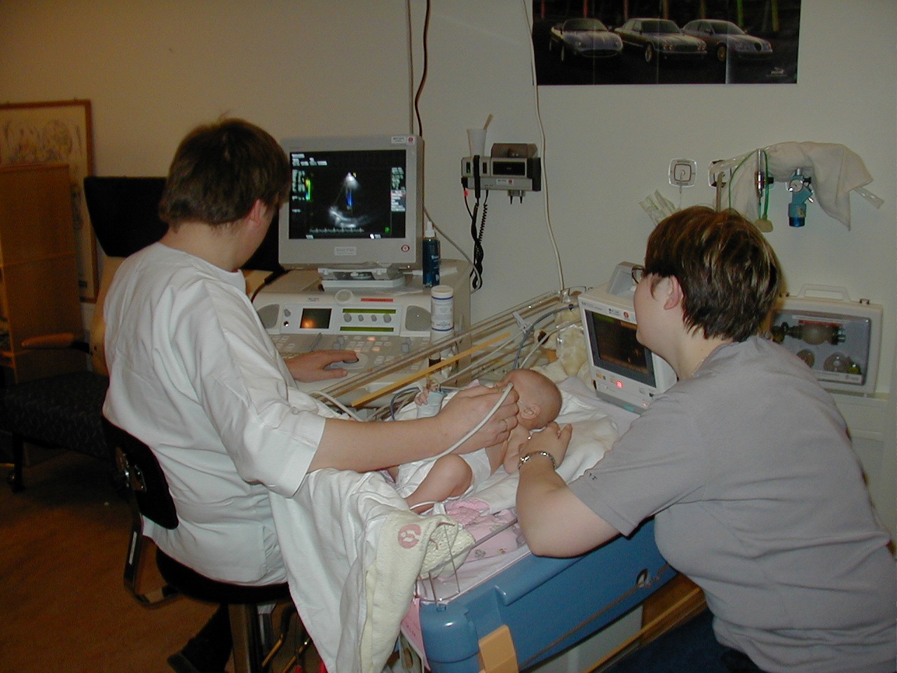
Images of Medical Ultrasonography

Ultrasound of carotid artery

B-flow image of venous reflux. Video is available.

Urinary bladder (black butterfly-like shape) and hyperplastic prostate (BPH) visualized by medical ultrasound

Intravascular ultrasound image of a coronary artery (left), with color-coding on the right, delineating the lumen (yellow), external elastic membrane (blue) and the atherosclerotic plaque burden (green)

Ultrasound of human heart showing the four chambers and mitral and tricuspid valves
![Orthogonal planes of a three-dimensional sonographic volume with transverse and coronal measurements for estimating fetal cranial volume[17][18]](https://upload.wikimedia.org/wikipedia/en/thumb/1/13/Head-3D.jpg/330px-Head-3D.jpg)
Orthogonal planes of a three-dimensional sonographic volume with transverse and coronal measurements for estimating fetal cranial volume[17][18]

Neck ultrasound

A modern medical ultrasound scanner

Curvilinear array transducer
Applications In Medicine
Ultrasonography has many important applications in medicine. It's commonly used during pregnancy to check how the baby is growing and if it is healthy. 👶It can also help diagnose conditions like gallstones, kidney stones, and heart problems. Cartilage and muscles can be examined using ultrasound as well. Doctors can spot cysts or tumors in organs quickly! 🦠Ultrasound also helps guide doctors during certain procedures, like injections. The cool thing is, since it's non-invasive, it feels safe and easy, making it a favorite for many doctors! 🏥
Types Of Ultrasonography
There are different types of ultrasonography! The most common types include 2D ultrasound, which gives flat images, and 3D ultrasound, which provides more detailed pictures. 🖼️ Then, there's 4D ultrasound that can show live moving images – like seeing the baby wave its tiny hand! 👋Transvaginal ultrasound is used for women, and it involves a small device inserted gently for better pictures. Doppler ultrasound allows doctors to see how blood flows in the body. 🚀Each type helps doctors in unique ways to understand our health better!
How Ultrasonography Works
Ultrasonography works by sending sound waves into the body through a special hand-held device called a transducer. 🎤When the sound waves bounce back, the device captures them and creates an image! ⭐️ The sound waves travel faster in solid parts like bones and slower in liquids such as blood. That's how doctors can tell what’s happening inside! The great thing is that it doesn't hurt at all! Patients just lie down while the doctor moves the transducer over the area being checked, and the results appear on a special screen. 📺
History Of Ultrasonography
Ultrasound started its journey in the 1950s. The first sonograms (ultrasound images) were created by a scientist named Dr. Ian Donald in Scotland! 🏴☠️ He used sound waves to look at babies inside their mothers. Since then, ultrasound has improved a lot. In the 1970s and 1980s, technology advanced, allowing us to see clearer pictures. The 3D and 4D ultrasound, which shows movement, emerged later! Today, hospitals use these machines to quickly check on patients and for many cool reasons, like finding out if someone is having a boy or girl! 🎀🎩
Case Studies In Ultrasonography
In recent case studies, doctors have used ultrasounds to find serious conditions early. For instance, a study showed that pregnant women who had routine ultrasounds could help detect issues, leading to healthier babies. 👶Another exciting case involved a patient whose leg pain turned out to be a blood clot, easily spotted during an ultrasound! 🚑Also, doctors found out that regular ultrasound exams helped in diagnosing heart problems earlier than later. These examples show how vital ultrasound is in helping doctors make important medical decisions! 📈
Common Procedures And Protocols
Many common procedures involve ultrasonography. For example, doctors perform abdominal ultrasounds to check organs like the liver and kidneys. A pelvic ultrasound helps assess women's reproductive health, while a fetal ultrasound checks the growth of babies. 🏋️♀️ The protocols usually involve having the patient lie down comfortably and applying gel to help the sound waves travel! Then, the ultrasound technician moves the transducer over the area to get images. This can take 30 minutes to an hour! They capture the images and send them to a doctor for review! 📊
Safety And Risks Of Ultrasonography
Ultrasonography is considered very safe with no risks related to radiation, unlike X-rays! 🥳However, it's important to only use it when necessary to make sure patients are healthy. Sometimes, the ultrasound machine can get a little chilly, making the gel feel cool on the skin. 🥶While there are no side effects, it's essential for trained professionals to use the equipment. If done properly, ultrasound has no harmful effects on the mother or the baby! This is part of why doctors love using this magical technology! 🌟
Future Of Ultrasonography Technology
The future of ultrasonography technology looks super bright! 🌈Scientists and engineers are working on making machines smaller, faster, and even better at making clear images. They are also trying out new ways to combine ultrasound with other technologies, like artificial intelligence. 🤖This can help doctors diagnose problems faster and more accurately. Imagine hand-held devices we can use at home to check our health! The use of 3D and 4D images will only get better, allowing excited parents to see their babies in motion! This means amazing things for healthcare ahead! 🚀
Ultrasonography Vs Other Imaging Techniques
Ultrasonography has unique advantages over other imaging techniques like X-rays and MRI scans. 🤔For example, while X-rays use radiation, ultrasounds use sound waves, making them safer for everyone! 🛡️ Also, ultrasounds are often quicker and can be done right at the doctor's office. MRI scans make loud noises and require a tunnel-like machine, while ultrasounds are quiet and gentle. However, MRIs provide even clearer pictures for some issues. It’s important to choose the right method! Doctors decide the best imaging technique based on what they are trying to find! 🤓
Training And Certification For Ultrasonographers
To become an ultrasonographer, you need special training! 🏫First, you usually need to finish high school, then go to college or a training program focusing on ultrasound. This often takes 1 to 4 years! 📚After that, students learn how to work the machines and understand human anatomy. Once they finish their education, they take an exam to become certified. This means they are officially allowed to work! 👍Ongoing education is important too, as they need to keep up with new technologies to provide the best care.
Medical Ultrasonography Quiz
Learn more about Medical Ultrasonography
