Atomic Force Microscopy Facts For Kids

Atomic force microscopy (AFM) is a high-resolution imaging technique that uses a mechanical probe to measure the forces between the probe and the sample surface, allowing for the visualization of surface structures at the nanoscale.

Select reading age
Go deeper More articles
Atomic Force Microscopy
Atomic Force Microscopy Facts For Kids
Videos & Quizzesincluded

About this topic

Atomic Force Microscopy (AFM) is a cool science tool that helps us see tiny things we can't see with our eyes! 🔭It was invented in 1986 by a smart scientist named Gerd Binnig. AFM looks at materials at an atomic level, which means it can see smaller than a single hair! 🧑‍🔬 It works by using a super sharp tip that "feels" the surface of the object, like how you feel a bumpy road when you touch it with your hand. AFM is used in laboratories all over the world, from countries like the USA to Japan! 🌍
Explore with ChatDino

Key Points

An AFM is made of several important parts!
AFM works like a tiny, very sensitive finger!
There are different ways to use an AFM!
AFM has many great advantages!
The future of AFM looks exciting!
Learning about AFM can be fun!
Preparing samples for AFM is very important!
AFM is used in many exciting fields!

Photos of Atomic Force Microscopy

An atomic force microscope on the left with controlling computer on the rightImage by Zureks, licensed under Creative Commons Attribution-Share Alike 3.0

An atomic force microscope on the left with controlling computer on the right

Fig. 3: Typical configuration of an AFM. (1): Cantilever, (2): Support for cantilever, (3): Piezoelectric element (to oscillate cantilever at its eigen frequency), (4): Tip (Fixed to open end of a cantilever, acts as the probe), (5): Detector of deflection and motion of the cantilever, (6): Sample to be measured by AFM, (7): xyz drive, (moves sample (6) and stage (8) in x, y, and z directions with respect to a tip apex (4)), and (8): Stage.Image by Tom Toyosaki, licensed under Creative Commons Attribution-Share Alike 4.0

Fig. 3: Typical configuration of an AFM. (1): Cantilever, (2): Support for cantilever, (3): Piezoelectric element (to oscillate cantilever at its eigen frequency), (4): Tip (Fixed to open end of a cantilever, acts as the probe), (5): Detector of deflection and motion of the cantilever, (6): Sample to be measured by AFM, (7): xyz drive, (moves sample (6) and stage (8) in x, y, and z directions with respect to a tip apex (4)), and (8): Stage.
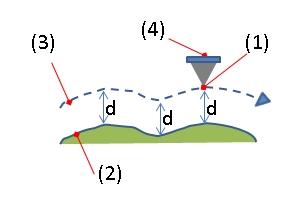
Fig. 5: Topographic image forming by AFM. (1): Tip apex, (2): Sample surface, (3): Z-orbit of Tip apex, (4): Cantilever.Image by Tom Toyosaki, licensed under Creative Commons Attribution-Share Alike 4.0

Fig. 5: Topographic image forming by AFM. (1): Tip apex, (2): Sample surface, (3): Z-orbit of Tip apex, (4): Cantilever.

Atomic Force Microscopy — photo 4Image by AFM_(used)_cantilever_in_Scanning_Electron_Microscope,_magnification_1000x.GIF : SecretDisc derivative work: Materialscientist ( talk ), licensed under Creative Commons Attribution-Share Alike 3.0

Atomic Force Microscopy — photo 4

Atomic Force Microscopy — photo 5Image by AFM_(used)_cantilever_in_Scanning_Electron_Microscope,_magnification_3000x.GIF : SecretDisc derivative work: Materialscientist ( talk ), licensed under Creative Commons Attribution-Share Alike 3.0

Atomic Force Microscopy — photo 5

Atomic force microscope topographical scan of a glass surface. The micro and nano-scale features of the glass can be observed, portraying the roughness of the material. The image space is (x,y,z) = (20 μm × 20 μm × 420 nm).

Atomic force microscope topographical scan of a glass surface. The micro and nano-scale features of the glass can be observed, portraying the roughness of the material. The image space is (x,y,z) = (20 μm × 20 μm × 420 nm).

Single polymer chains (0.4 nm thick) recorded in a tapping mode under aqueous media with different pH.[11]Image by Yurko, licensed under Creative Commons Attribution-Share Alike 3.0

Single polymer chains (0.4 nm thick) recorded in a tapping mode under aqueous media with different pH.[11]

Model for AFM water meniscusImage by Ilaria.denti, licensed under Creative Commons Attribution-Share Alike 4.0

Model for AFM water meniscus

AFM beam-deflection detectionImage by Creepin475, licensed under Creative Commons Attribution-Share Alike 3.0

AFM beam-deflection detection

🧩 Components Of An Afm

An AFM is made of several important parts! 🧩First, there's the cantilever, which holds the sharp tip. Next, we have the scanner, which moves the tip precisely over the surface. The laser light measures how much the cantilever bends. 💡There’s also a computer system that creates images and stores data! The whole setup usually sits on a stable table to avoid shaking, which can mess up the measurements. Last, there's a special holder for the samples we want to study, making everything fit perfectly! 🎯
Go deeper

🤏 Principle Of Operation

AFM works like a tiny, very sensitive finger! 🤏When the sharp tip touches the surface of an object, it moves up and down depending on the bumps and grooves. This movement is recorded by a computer, turning what it sees into a picture! 🌈The tip is attached to a tiny arm called a cantilever. If the tip gets closer to the object, it feels a pull, and the cantilever bends. The amount of bending helps scientists learn about the object's shape and texture. It's like magic! 🪄
Go deeper

🤓 Types Of Afm Techniques

There are different ways to use an AFM! 🤓For example, in "Contact Mode," the tip lightly touches the surface to feel the texture. In "Tapping Mode," the tip bounces up and down instead of staying in contact, which minimizes damage to soft items. Another method is "Non-Contact Mode," where the tip never touches the surface but detects tiny forces! 🪶Each technique is helpful for studying different materials, like metals, plastics, or even living cells! 🌱Scientists choose the technique based on what they want to discover!
Go deeper

🎊 Advantages And Limitations

AFM has many great advantages! 🎊It can see objects as small as 1 nanometer, which is one billionth of a meter! 🥳It’s also super precise and can handle different surfaces. However, it has limits too! 🛑For example, AFM can be slow and can struggle with very large objects. Some materials might also damage the tip if they are too hard. Scientists need to think carefully about using AFM, but it gives amazing details that other tools can't! 🥇
Go deeper

🤩 Future Developments In Afm

The future of AFM looks exciting! 🤩Scientists are working on making AFM faster and able to see even smaller things, like individual atoms! 🧬Researchers are also combining AFM with other technologies, like with lasers for even better pictures! 🌟In the future, we might be able to study living cells without hurting them! This could change medicine and science forever! As technology improves, we can only imagine what new discoveries await us and how AFM will help! 🚀
Go deeper

📚 Education And Resources On Afm

Learning about AFM can be fun! 📚Many universities and science centers offer workshops and classes about microscopy. Websites like NASA and National Geographic have cool articles about it too! 🌌If you want to experiment, ask your teachers if your school has a science lab! 🏫There, you can see how AFM works in action! You can also watch videos about AFM on platforms like YouTube. Keep exploring, because science is full of amazing things just waiting for you! 🔍
Go deeper

🧪 Sample Preparation And Imaging

Preparing samples for AFM is very important! 🧪Objects must be clean and flat. If they're not, the images won't be good! Sometimes, scientists coat samples with special materials to help the AFM tip get better readings. 📏The surface also needs to be dry—no water! After preparing, the sample is placed in the AFM holder, and the fun begins! The team can start taking images, creating 3D pictures, and learning new things about the sample! 🖼️
Go deeper

⚗️ Applications Of Atomic Force Microscopy

AFM is used in many exciting fields! ⚗️ In materials science, it helps engineers create better gadgets, like phones and computers. 🎮In biology, AFM can examine tiny parts of cells, helping researchers learn about diseases! 🌼The food industry uses AFM to test food safety! 🍎Even in art, AFM helps restorers understand how to fix ancient paintings! 🎨With AFM, scientists around the world can explore everything from technology to medicine.
Go deeper

🧠 Take the Quiz about Atomic Force Microscopy

Question 1 of 10

What does AFM stand for?

Question 2 of 10

What type of cantilever is used in AFM?

Question 3 of 10

In which mode does AFM typically operate to avoid damaging soft samples?

Question 4 of 10

What type of forces are primarily measured in AFM?

Question 5 of 10

What kind of samples can AFM analyze?

Question 6 of 10

What is a key advantage of AFM compared to electron microscopy?

Question 7 of 10

What is the typical resolution that AFM can achieve?

Question 8 of 10

Which application would NOT typically use atomic force microscopy?

Question 9 of 10

What type of technology can be used to enhance the imaging capabilities of AFM?

Question 10 of 10

What type of data can AFM provide besides topographical images?

🔒

Your results

Answer all 10 questions to see how you did.

Kid-safe AI powered by AstroSafe

DIY.org

Kid-safe AI for discovery and learning. Ask anything, explore everything, learn with confidence.

Product

  • Pricing
  • App
  • All Apps

Resources

  • Blog
  • Kids Wikipedia
  • Worksheets
  • Activities for Kids
  • Build a Hacker Simulator
  • SafeTube

Company

  • About
  • Terms & Conditions
  • Privacy Policy
  • AstroSafe
TwitterInstagramYouTube

© 2026 DIY.org. All rights reserved. · Powered by AstroSafe